Measuring the Body in Orbit with Ultrasound · Stellar Amenities Lab
[ The science ahead ]

The Human Body Is the Next Frontier of Science

Launch, stations and landers have been remade in a decade. Our understanding of what happens to the body in space has barely moved in sixty years, and that is where the next breakthroughs are.

Spaceflight is the fastest experiment in human biology we have. Bone that takes a decade to thin on Earth thins in months in orbit. Arteries stiffen, muscle fades and sleep breaks down on a timescale a laboratory can actually follow. Every crew member who flies is showing us, in compressed form, processes that shape every life on the ground.

We have mostly looked away. Measurements are taken before launch and after landing, and the months in between, where the change actually happens, go largely unrecorded. The science is waiting on the instruments.

The world ahead

Within the next two decades, more than a hundred people may be living off Earth at any one time, on commercial stations and on the Moon. Each of them will carry a continuous record of how their body adapts. Pooled across crews and missions, that record becomes the largest longitudinal study of human ageing and recovery ever assembled.

It will change how missions are planned, how habitats are designed and how countermeasures are tested. And it will come home: what protects an astronaut for six months is close to what protects a patient on bed rest, and what reverses decline in orbit is worth knowing everywhere.

“We used to wait until they landed to find out.”

It starts with an instrument a crew can wear. Read our technical approach.

Technical note · October 2026

Measuring the body in orbit with ultrasound

Bone, muscle, blood vessels and the eye all change within weeks of leaving Earth. Most of what we know about those changes comes from measurements taken before launch and after landing. We are building an ultrasound system that a crew wears instead of operates, so the record runs through the mission itself.

Why ultrasound

A habitat has no room for MRI or CT. Both are large, heavy and power-hungry, and CT adds radiation to a crew already exposed to more of it than anyone on Earth. Ultrasound is the only imaging method that fits in a habitat, runs on modest power and is safe to repeat daily.

It has flown before. Crews on the International Space Station have used ultrasound for years, and the eye and the jugular vein are two of the places it has shown changes that ground-based tests missed. What has changed is the hardware. Chip-based probes, of the kind Butterfly Network makes, have brought a scanner that once filled a cart down to the size of a phone.

The operator problem

On the station today, a crew member holds the probe on their own body while a sonographer on the ground guides them by voice. The result depends on how well the probe is placed and held, and every session costs crew time. That limits ultrasound to occasional checks, and occasional checks cannot show how a change develops.

Our system removes the operator. The probe is worn, positions itself against fixed landmarks on the body, and runs the same scan the same way every session. Consistency matters more than any single image: two scans are only comparable if they were taken the same way.

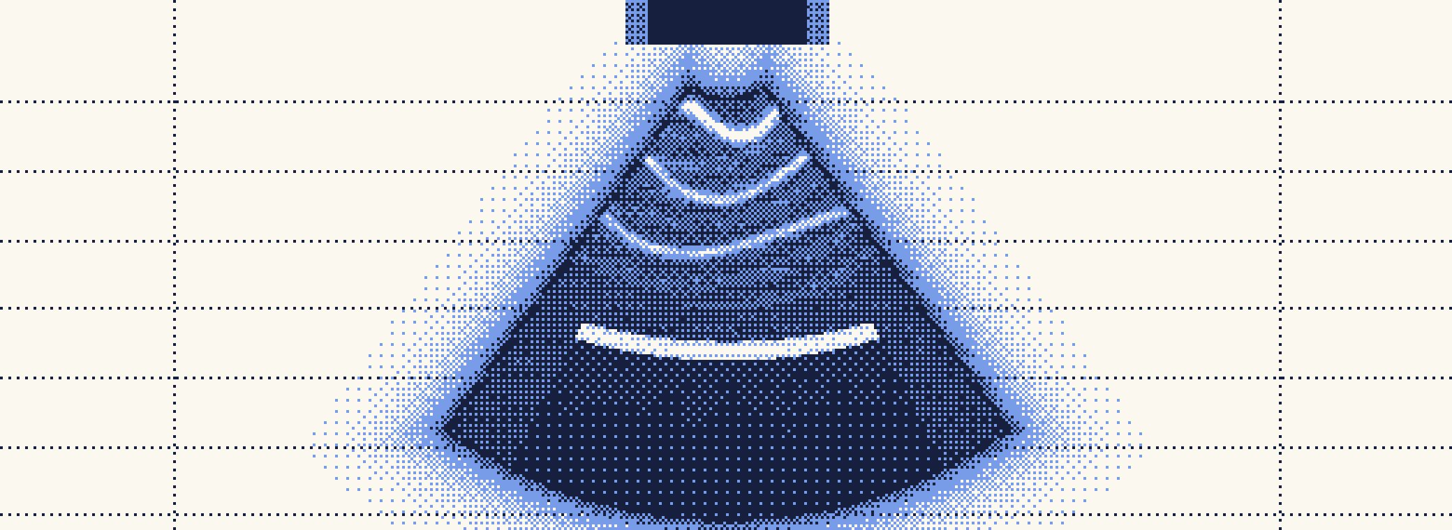
one handheld scan A snapshot placement varies · signal blurs
{{ sessionLabel }} A record
Illustrative. A single scan taken by hand varies with how the probe is placed. Scans taken the same way, session after session, stack into a record where structure and change become visible. Drag the slider to add sessions.

What we measure

BoneQuantitative ultrasound at the heel and shin, tracking the loss of 1 to 1.5 percent a month that crews see in weight-bearing bone.
MuscleThickness and cross-section of the calf and thigh, the muscles that waste fastest without load.
VesselsCarotid stiffness and jugular flow. Six months in orbit can stiffen the carotid as much as ten to twenty years of ageing on Earth.
EyeOptic nerve sheath diameter, an ultrasound marker for the eye changes that affect many long-duration crew.

From images to a model

A probe produces far more raw signal than a clinical image shows. Most of it is discarded by hand-tuned processing before anyone looks at it. We keep the raw data and the conditions it was taken under, so that models trained on the record can recover more than today's pipelines do, and so that each measurement can be compared with every one before it.

The record pairs with strength and loading data from training hardware on the same crew. That pairing is what links a change in bone or muscle to what the body was actually doing.

Where this stands

The wearable form and the auto-scanning software are in development in the Lab. The first flight test is a parabolic campaign in late October 2026, to check that placement and image quality hold through changing gravity. A demonstration on a commercial station is the next step.

Notes The figure above is illustrative and does not show measured data. Carotid stiffness: Hughson et al., American Journal of Physiology, 2016. Bone loss: NASA Human Research Program evidence report.